Digital radiography machines have become the cornerstone of modern medical imaging. This technology surpasses traditional film-based systems by delivering faster results, enhanced image quality, and streamlined workflows. From mobile digital radiography units deployed in emergency settings to advanced digital dental x ray machines used in dental clinics, these devices revolutionize diagnostic capabilities across healthcare facilities.
You interact daily with various forms of medical imaging equipment, including handheld ultrasound on phone solutions and full-scale DR X-ray systems integrated within hospital networks. Each device demands meticulous setup to ensure patient safety and image accuracy. Improper configuration can lead to repeated exposures, diagnostic errors, and increased radiation risks.
This article serves as your comprehensive, field-ready checklist for setting up a digital radiography machine safely and effectively. Whether you are a radiology devices manager responsible for equipment readiness or a technologist operating a portable diagnostic imaging unit, this guide covers essential steps from initial preparation through post-examination review. Following these protocols helps optimize image quality while minimizing radiation exposure—critical goals in today’s healthcare environment.
In addition to these practical steps, it's also important to consider the role of effective documentation in the setup and operation of these machines. Creating effective documentation can greatly enhance the efficiency of your processes.
Moreover, to maximize the benefits of your documentation, it's essential to adopt certain strategies that streamline its use. Investing in online documentation can also provide significant advantages, such as easier access and better organization of information. Finally, don't underestimate the hidden benefits of investing in good documentation, which can lead to improved patient outcomes and more efficient workflows.
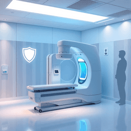
Understanding Digital Radiography Machines
Digital radiography machines represent a significant advancement over traditional film-based systems. Instead of capturing images on physical film, these machines use digital detectors to convert X-rays into electronic signals. This produces immediate high-resolution images that can be viewed, enhanced, and stored digitally. The elimination of chemical processing speeds up workflows and reduces environmental impact.
Key Differences from Traditional Film-Based Systems
- Image Capture: Digital radiography equipment uses flat-panel detectors or charged-coupled devices (CCDs) rather than film.
- Image Availability: Instant preview and the ability to manipulate images improve diagnostic accuracy.
- Storage and Sharing: Images are easily archived in Picture Archiving and Communication Systems (PACS) and shared electronically.
- Radiation Dose: Optimized exposure settings often reduce patient radiation dose compared to film methods.
Types of Digital Radiography Machines
Portable radiography machines offer flexibility for bedside imaging or remote locations. These compact units are battery-powered, lightweight, and designed for quick setup, making them ideal for emergency rooms, intensive care units, or field hospitals.
Mobile X-ray machines are larger than portable devices but still designed for mobility within hospital facilities. Mounted on wheels with adjustable arms, they provide more power and advanced features while maintaining transportability.
Fixed digital radiography equipment installed in radiology departments provides the highest image quality for routine examinations. These systems integrate sophisticated detectors, automated positioning aids, and advanced software algorithms.
Computed Radiography Machines in Diagnostic Imaging
Computed radiography (CR) machines act as a bridge between analog film systems and fully digital radiography. They use photostimulable phosphor plates to capture X-ray images. After exposure, these plates are scanned by a CR reader to digitize the image.
CR systems remain valuable where full digital upgrades haven't been implemented yet or as backup solutions. Their adaptability allows integration with existing X-ray generators and compatibility with various patient positioning setups.
Role in Hospital Imaging Equipment Setups
Digital radiography equipment forms the backbone of diagnostic imaging services in modern healthcare facilities:
- Supports diverse clinical needs from trauma assessment to dental evaluations using specialized dental X-ray equipment.
- Enhances workflow efficiency by reducing image acquisition time and facilitating rapid diagnosis.
- Enables compliance with radiation safety protocols through dose monitoring capabilities.
- Facilitates multidisciplinary collaboration by providing clear images accessible across hospital networks.
Integration of mobile and portable devices extends imaging capabilities beyond fixed rooms, crucial for critically ill patients or surgical suites requiring immediate imaging support.
Radiographic testing equipment used outside medical contexts shares some technology but focuses on industrial applications such as non-destructive testing. Understanding distinctions helps maintain focus on medical imaging device requirements.
The variety of digital radiography machines available allows tailoring solutions to specific clinical environments while maintaining consistent image quality standards essential for accurate diagnosis.
In this context, the importance of comprehensive user guides cannot be overstated. Such documentation plays a crucial role in ensuring that medical professionals can effectively utilize the advanced features of digital radiography machines. It's essential to understand the benefits of user guides in this regard.
Moreover, it's vital to recognize that poor documentation practices can significantly hinder the effective use of these machines. Why your documentation is killing your should be a key consideration when implementing new technology in medical settings.
Pre-Examination Setup Checklist
Radiation safety and patient accuracy begin before the first exposure. This checklist ensures you address key elements to protect patients and staff, and optimize exam quality.
Patient Identification
Confirm the patient's identity using at least two unique identifiers. Commonly used identifiers include:
- Full name
- Date of birth
Verifying identity prevents mix-ups that could lead to incorrect imaging or unnecessary radiation exposure. Cross-check these details with the exam order and patient wristband when available.
Appropriateness of Requested Exam
Evaluate whether the requested radiographic exam aligns with the clinical indication. This step avoids unnecessary radiation by:
- Confirming the exam is justified by current symptoms or diagnosis
- Consulting with referring physicians if clarification is needed
- Considering alternative imaging modalities without ionizing radiation if appropriate
Justification ensures radiation exposure is medically warranted, adhering to ALARA (As Low As Reasonably Achievable) principles.
Pregnancy Status Verification
For females of childbearing age, confirming pregnancy status is critical to minimize fetal radiation risk. Steps include:
- Asking the patient directly about pregnancy or recent menstruation
- Reviewing medical records for documented pregnancy tests
- Utilizing urine or blood pregnancy testing if required by protocol or uncertainty exists
Documenting pregnancy status guides decision-making on exam necessity, shielding use, or postponement.
Radiation Monitoring Devices for Staff Safety
Staff protection depends on consistent use of radiation monitoring devices, such as film badges or electronic dosimeters. Responsibilities include:
- Ensuring all personnel wear their assigned badges properly during procedures
- Checking device functionality before shifts
- Regularly reviewing dose reports to identify trends or overexposures
Radiation monitoring devices are integral to maintaining safe occupational exposures within regulatory limits.
Equipment and Environment Preparation
Prepare your digital radiography machine and examination space by:
- Inspecting equipment for damage or malfunction (e.g., cables, detectors)
- Verifying calibration and proper functioning of image receptors
- Ensuring availability of necessary accessories like grids, markers, and shields
- Setting up clean and organized patient positioning aids
- Confirming room cleanliness and removal of any unnecessary items that could interfere with workflow or cause contamination
Adequate preparation reduces delays, prevents errors, and supports smooth operation during image acquisition.
This pre-examination setup creates a controlled environment prioritizing patient safety and optimal image quality. Next steps focus on verifying equipment readiness and applying safety measures tailored to each imaging procedure.
Equipment Preparation and Safety Checks
Setting up your digital radiography machine requires careful attention to both equipment functionality and radiation safety measures. Ensuring optimal performance while minimizing patient and staff exposure requires following key preparation steps.
Inspecting the Digital Radiography Machine Functionality
Before any patient examination, conduct a thorough inspection of the diagnostic imaging device:
- Power and Connectivity: Verify that the machine powers on without error messages and that all cables and wireless connections are secure.
- Image Receptor Integrity: Check the detector for any physical damage, dust, or artifacts that could degrade image quality.
- Software and Calibration: Confirm the latest software version is installed; run calibration protocols to ensure accurate image capture parameters.
- Mechanical Components: Test movement of the X-ray tube arm, collimator blades, and positioning locks to guarantee smooth operation.
A malfunctioning radiology device compromises image quality and may increase repeat exposures, undermining radiation safety principles.
Proper Use and Selection of Anti-Scatter Grids
Anti-scatter grids play a crucial role in enhancing image contrast by absorbing scattered radiation before it reaches the image receptor. Their application should be tailored according to body part thickness:
- Use grids for body parts thicker than approximately 10 cm (e.g., abdomen, pelvis) to reduce scatter effectively. For more information about grids, you can refer to this resource.
- Avoid grids on thinner areas like extremities or pediatric patients, as they increase dose without significant benefit.
- Select a grid ratio and frequency appropriate for the examination; higher grid ratios improve scatter rejection but require dose adjustments.
Appropriate grid use balances improved diagnostic image quality with minimized radiation exposure.
Collimating the Primary Beam
Beam collimation limits radiation to only the anatomical area of interest. Precise collimation reduces irradiated tissue volume and scatter radiation reaching the detector:
- Adjust collimator blades so the X-ray field matches or slightly exceeds the size of the targeted anatomy.
- Avoid excessively large fields, which unnecessarily expose surrounding tissues.
- Mark collimation settings on images when feasible for documentation.
Effective collimation optimizes patient safety by controlling dose distribution while maintaining necessary diagnostic information.
Applying Appropriate Shielding
Shielding serves as an additional protective measure, especially important when sensitive organs lie near or within the X-ray field:
- Use gonadal shields whenever reproductive organs are within or adjacent to the primary beam path unless it compromises diagnostic information.
- Apply thyroid collars or breast shields selectively based on exam type and clinical guidelines.
- Ensure shielding devices are properly positioned without obscuring anatomy critical for diagnosis.
Radiology device managers must enforce strict shielding protocols alongside equipment checks to maintain compliance with radiation protection standards.
Preparation steps focusing on equipment performance combined with precise control of radiation exposure factors form a foundation for safe digital radiography practice. Adhering to these procedures protects patients and staff while supporting high-quality imaging outcomes.
Patient Positioning, Immobilization, and Technical Parameter Settings for Optimal Imaging
Patient positioning is crucial for obtaining high-quality diagnostic images while minimizing radiation exposure. Each type of examination requires specific positioning techniques that align the anatomical area of interest with the image receptor and primary beam. Knowing the standard positioning protocols ensures accurate representation of anatomy and reduces the need for repeat exposures.
Key aspects of patient positioning include:
- Aligning the body part parallel or perpendicular to the image receptor depending on the exam type.
- Ensuring the central ray is directed precisely through the targeted anatomy to avoid distortion.
- Adjusting patient posture to open joint spaces or highlight structures critical for diagnosis.
Immobilization Techniques
Immobilization techniques help prevent motion artifacts that degrade image quality. Any movement during exposure can blur anatomical details, reducing diagnostic value and potentially leading to additional imaging sessions. Immobilization methods vary from simple verbal instructions to physical devices such as foam pads, sandbags, straps, or specialized immobilizers.
Effective immobilization involves:
- Using support devices tailored to the patient's size and exam requirements.
- Applying gentle but firm restraint without causing discomfort or anxiety.
- Collaborating with patients by explaining the importance of remaining still during exposure.
Clear communication enhances patient cooperation and contributes significantly to successful imaging outcomes. Explaining breathing instructions—such as holding breath or breathing shallowly—reduces involuntary motion affecting thoracic or abdominal images. Technologists should provide concise directions before each exposure and confirm patient understanding.
Technical Parameter Selection
Selecting technical parameters like kilovoltage peak (kVp) and milliampere-seconds (mAs) depends on body part thickness, composition, and diagnostic goals. Appropriate kVp settings improve penetration through tissues, while correct mAs controls radiation dose and image brightness. Balancing these parameters optimizes contrast resolution and signal-to-noise ratio without unnecessary patient dose.
Guidelines for technical parameter selection include:
- Increasing kVp for thicker or denser body parts (e.g., pelvis) to ensure adequate photon penetration.
- Adjusting mAs proportionally with kVp changes to maintain consistent image receptor exposure.
- Applying manufacturer-recommended settings based on device-specific calibration charts.
- Utilizing automatic exposure control (AEC) features when available to standardize exposures.
Implementing precise patient positioning combined with effective immobilization and tailored technical settings elevates image quality substantially. These practices minimize artifacts, reduce repeat rates, and enhance diagnostic confidence when using diagnostic radiography equipment in clinical environments.
Image Acquisition Process, Post-Acquisition Review, Quality Control, and Workflow Pause Points for Safety Assurance
The image acquisition phase demands precision to achieve diagnostic-quality images while maintaining radiation protection protocols. Proper alignment between the primary beam and the image receptor is fundamental. Misalignment can cause geometric distortions, image cut-off, or repeats, unnecessarily increasing patient dose. This applies critically in portable diagnostic imaging, where space constraints challenge setup accuracy.
Key steps to ensure correct alignment:
- Position the image receptor parallel to the anatomical area of interest.
- Align the central ray of the x-ray beam perpendicular to both the receptor and body part.
- Verify source-to-image distance (SID) settings are optimal for exam type.
Placement of anatomical markers is mandatory on all images. These markers denote laterality (right/left), patient position, and orientation critical for accurate interpretation by radiologists. Markers should be placed outside the anatomy but within the collimated field to avoid obscuring diagnostic details.
After exposure, immediate review of displayed images on digital monitors allows technologists to verify:
- Image clarity and absence of motion artifacts.
- Correct anatomical coverage.
- Patient identification matches requested exam.
- Proper marker placement.
This real-time assessment helps decide if a repeat is necessary before moving on, reducing retakes and radiation dose.
Incorporating workflow pause points serves as safety checkpoints throughout the imaging process:
- Pre-exposure pause: Confirm patient ID, exam appropriateness, positioning, alignment, shielding application, and technical factors.
- Post-exposure pause: Review image quality and documentation completeness.
- Pre-patient release pause: Ensure no additional views are needed and that all safety measures were followed.
Pause points empower technologists to catch errors early, enforce radiation protection standards consistently, and enhance overall workflow efficiency.
Radiation protection remains integral during image acquisition. Collimation limits primary beam size to target anatomy only. Appropriate shielding devices like gonadal shields must be applied when indicated without compromising image quality or risking repeats.
Portable diagnostic imaging environments demand extra vigilance with image receptor alignment and workflow discipline due to variable conditions outside controlled radiology suites. Utilizing this checklist-driven approach ensures high-quality imaging with minimized radiation risk in any setting.
Specialized Considerations in Radiology Device Management
Radiology device manager roles extend beyond equipment handling to include specific protocols for safely imaging patients with implanted cardiac devices. These devices require tailored approaches to prevent interference and ensure patient safety.
Pacemaker Radiology Precautions
- Device Identification: Confirm presence and type of pacemaker before imaging. Obtain details from patient records or device identification cards.
- Imaging Modality Selection: Digital radiography is generally safe, but avoid MRI unless the device is MRI-compatible.
- Radiation Exposure: Limit exposure near the pacemaker site to reduce risk of device malfunction.
- Positioning: Avoid placing the primary beam directly over the pacemaker if possible.
- Monitoring: Continuous cardiac monitoring may be necessary during lengthy or complex procedures.
- Communication: Inform the radiology assistant or cardiac devices specialist when a pacemaker is involved for additional support.
Loop Recorder Radiology Guidelines
- Device Recognition: Loop recorders are small, subcutaneous devices used for long-term cardiac rhythm monitoring.
- Imaging Safety: Routine digital radiography is safe; however, inform staff of device location to avoid beam overlap.
- Interference Prevention: Avoid use of strong electromagnetic fields or prolonged exposure near the device.
- Documentation: Note device presence clearly in imaging reports and patient records. Implementing a robust document management system can streamline this process.
Impella Device Radiology Protocols
The Impella device, a percutaneous ventricular assist device, introduces unique challenges in diagnostic imaging.
- Pre-Imaging Assessment: Verify Impella presence with clinical teams before proceeding.
- Device Positioning Awareness: Ensure awareness of catheter and motor pump position to avoid dislodgement during positioning.
- Radiation Dose Management: Minimize radiation dose near the device to prevent damage or interference with function.
- Image Acquisition Modifications: Adjust technique factors to compensate for metallic components causing artifacts.
- Emergency Preparedness: Have protocols ready for immediate response in case of device-related complications during imaging.
Role of Radiology Devices Manager and Assistant Cardiac Devices Specialists
Managing radiology devices involves coordination with specialists trained in cardiac implantable electronic devices (CIEDs). Their expertise supports:
- Safe integration of digital radiography machines with implanted devices.
- Training technicians on pacemaker radiology precautions, loop recorder radiology guidelines, and impella device radiology protocols.
- Development and updating of institutional policies that reflect evolving best practices in imaging patients with cardiac devices.
Adhering to these specialized considerations ensures diagnostic quality while safeguarding sensitive cardiac devices during digital radiography procedures.
Conclusion
Ensuring a safe setup digital radiography machine is more than just a procedural step—it's a critical commitment to patient and staff safety. The importance of the field-ready checklist cannot be overstated, as it standardizes practices that directly impact image quality and radiation exposure. Following this checklist helps you:
- Optimize diagnostic image quality by ensuring correct equipment settings and patient positioning.
- Minimize radiation risks through appropriate use of shielding, collimation, and technical parameters.
- Reduce errors and repeat examinations, preserving valuable time and resources in busy clinical environments.
As a radiology devices manager or technologist working with hospital imaging equipment, continuous training remains essential. Medical imaging technology evolves rapidly, and staying up-to-date with the latest best practices enhances your ability to manage devices safely and efficiently. Adherence to evolving protocols strengthens quality assurance programs and supports compliance with regulatory standards.
Commitment to these principles fosters an environment where patient care excellence aligns with operational safety. Use this comprehensive guide as a foundation for establishing robust workflows that safeguard everyone involved in the diagnostic process while maximizing the performance of your digital radiography systems. Consistency in preparation, execution, and review transforms routine exams into reliable diagnostic tools that clinicians trust.
FAQs (Frequently Asked Questions)
What is a digital radiography machine, and how does it differ from traditional film-based systems?
A digital radiography machine is an advanced medical imaging device that captures X-ray images digitally, allowing for immediate image viewing and enhanced image processing. Unlike traditional film-based systems, digital radiography eliminates the need for chemical processing, reduces radiation exposure, and improves workflow efficiency in diagnostic radiology.
What are the essential pre-examination setup steps to ensure safety and accuracy in digital radiography?
Key pre-examination steps include verifying patient identity using at least two identifiers such as name and date of birth, confirming the appropriateness of the requested exam to avoid unnecessary radiation exposure, checking pregnancy status in females of childbearing age for radiation safety, ensuring staff use radiation monitoring devices like film badges properly, and preparing both equipment and environment adequately before examination.
How can equipment preparation and safety checks optimize image quality while minimizing radiation risks?
Proper equipment preparation involves inspecting the digital radiography machine's functionality before use, selecting appropriate anti-scatter grids based on body part thickness to enhance image quality, collimating the primary beam to limit radiation exposure strictly to necessary anatomical areas, and applying suitable shielding such as gonadal shields when applicable. These measures collectively optimize diagnostic accuracy while ensuring patient and staff safety.
What techniques are recommended for patient positioning and immobilization during digital radiography exams?
Accurate patient positioning tailored to specific examinations is crucial for optimal imaging. The use of immobilization devices or methods helps reduce motion artifacts. Clear communication with patients about breathing instructions and the importance of remaining still during exposure enhances image clarity. Additionally, selecting appropriate technical parameters like kilovoltage peak (kVp) and milliampere-seconds (mAs) based on body part thickness further improves image quality.
What are critical steps during image acquisition and post-acquisition review to maintain quality control in digital radiography?
Critical steps include ensuring proper alignment between the primary X-ray beam and the image receptor to prevent distortions or repeat exposures, placing anatomical markers correctly on images for accurate interpretation, immediately reviewing displayed images for clarity and correct patient identification details, and integrating workflow pause points to confirm completion of essential safety procedures before proceeding to subsequent stages.
What specialized precautions should be taken when imaging patients with cardiac devices such as pacemakers or Impella devices?
Special protocols must be followed when imaging patients with cardiac devices like pacemakers, loop recorders, or Impella devices. These include adhering to device-specific radiology guidelines to prevent interference or damage during diagnostic imaging. Radiology device managers should ensure all staff are trained on these precautions to maintain patient safety while achieving accurate diagnostic results.
Comments
Post a Comment